The epidemiology of muskuloskeletal disease Molecules, structures, patients and society
Musculoskeletal diseases are a rapidly growing cause of disability, chronic pain, and reduced quality of life in our ageing populations worldwide. Our aims are to gain novel insights into these diseases - their etiology, occurrence, natural history, treatments, prediction, disease monitoring, and disease burden - to allow for better healthcare decision-making and disease prevention.
To accomplish these goals, we use a multidisciplinary approach. For research on osteoarthritis, a chronic degenerative joint disease, we use human tissue biobanking, proteomics, and MR imaging to characterize the molecular and structural aspects of tissue degradation associated with the earliest stages of the disease. We are especially interested in the role of the meniscus in early knee osteoarthritis. Further, in population-based epidemiologic and health economic studies of musculoskeletal disease, we use physician-coded healthcare data from Sweden to understand the impact of these diseases on patients and society. Our previous work has contributed to improved understanding of musculoskeletal disease and impactful changes in their clinical management. To learn more, read about our projects.
Magnusson K, Turkiewicz A, Dell'Isola A, Englund M. 2024. Nature Communications.
Osteoarthritis is one of the most common musculoskeletal diseases and increases the risk of severe cardiovascular disease, like heart attack and stroke.
In some individuals, osteoarthritis and cardiovascular disease will co-occur.
This co-occurrence might be due to shared risk factors, for example high age, lifestyle factors and/or a shared genetic liability for the two diseases.
Here, we show that the correlation between osteoarthritis and cardiovascular disease can be explained by shared genetic factors, independent of high age and body weight, and also likely independent of lifestyle factors, like smoking and physical activity level.
Findings suggest that genetic factors that are shared for osteoarthritis and cardiovascular disease may contribute to both diseases.
Thus, the prevailing idea that osteoarthritis is predominantly a risk factor for cardiovascular disease is challenged. Our findings imply that the current diagnostic boundaries between these diseases may need to be re-evaluated.
Turkiewicz A, Magnusson K, Timpka S, Kiadaliri A, Dell'Isola A, Englund M. 2025. PLoS Medicine.
Health and lifestyle factors in youth is associated with cardiovascular or respiratory disease in adulthood, but associations with musculoskeletal disease is largely unknown.
We followed 40,000 Swedish males from 18-60 years old, with exposures of interest including physical health (e.g. body mass and height and muscle strength), self-reported lifestyle and health (e.g. smoking, alcohol and overall health).
We used the Swedish National Patient Register for incidence of common musculoskeletal, (including osteoarthritis), cardiovascular and respiratory diseases.
In conclusion we found that high body mass was a risk factor for all 3 studied groups of diseases, high cardiorespiratory fitness and high muscle strength in youth were associated with increased risk of musculoskeletal disease in middle age. We speculate that these associations are mediated by chronic overload or acute trauma.
Sillanpää N, Iivanainen M, Turkiewicz A, Sihvonen R, Paavola M, Taimela S, Järvinen TLN, Englund M. 2025. Osteoarthritis Cartilage.
We assessed 5-year effects of arthroscopic partial meniscectomy (APM) vs. placebo surgery on the development of the structural changes of the knee by MRI.
146 adults (mean age 52 years, range 35 to 65) was randomised with subjects having symptoms of degenerative medial meniscus tear, a tear verified in MRI and arthroscopy, and no advanced osteoarthritis at baseline.
We compared baseline and 5-year follow-up MRI, analysing progression of structural cartilage changes per subregion as main outcome, and used Osteoarthritis Knee Score scoring to derive subregional data on cartilage damage, osteophytes and bone marrow lesions as secondary outcomes.
In conclusion we found a slightly greater risk for progression of osteophytes in the APM group compared to the placebo-surgery group at 5 years after surgery.
Henriksen M, Runhaar J, Turkiewicz A, Englund M. 2024. Osteoarthritis Cartilage.
Exercise is universally recommended as a primary strategy for pain management in knee osteoarthritis (OA), which is based on results from more than 100 randomized controlled trials, comparing exercise to no-attention control groups.
However, due to difficulties with adequate placebo control, participant blinding and the use of patient-reported outcomes, the existing trials evidence is imperfect.
Here, we examined the existing evidence by providing a framework for assessment of possible causal relationships, which includes 9 considerations: Strength of association, Consistency, Specificity, Temporality, Biological Gradient (Dose-Response), Plausibility, Coherence, Experiment, and Analogy.
We conclude that the current evidence is not sufficient to support claims about (lack of) causality.
With our review, we hope to advance the continued global conversation about how to improve the evidence-based management of patients with knee OA.
B Wrammerfors ET, Törnquist E, Pierantoni M, Sjögren A, Tengattini A, Kaestner A, Zandt RI', Englund M, Isaksson H. 2024. Osteoarthritis Cartilage.
We investigated the feasibility of using neutron tomography to gain new knowledge of human articular cartilage degeneration in osteoarthritis (OA).
Different sample preparation techniques were evaluated to identify maximum intra-tissue contrast.
We used human articular cartilage samples from 14 deceased donors (18-75 years, 9 males, 5 females) and 4 patients undergoing total knee replacement due to known OA (all female, 61-75 years).
Neutron tomographic imaging was performed at two large scale facilities and the 3D images were evaluated for gradients in hydrogen attenuation as well as compared to images from absorption X-ray tomography, magnetic resonance imaging, and histology.
We concluded that neutron tomography is a viable technique for specialized studies of cartilage, particularly for quantifying properties relating to the hydrogen density of the tissue matrix or water movement in the tissue.
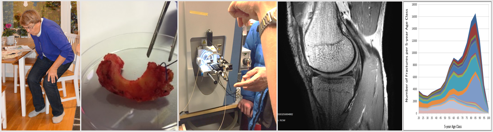
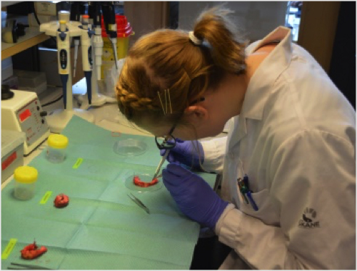
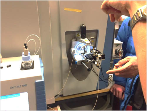
Molecular pathogenesis We perform biobanking from orthopaedic surgeries to target better understanding of osteoarthritis aetiology and pathogenesis using proteomics, towards identification of novel biomarkers of the disease.
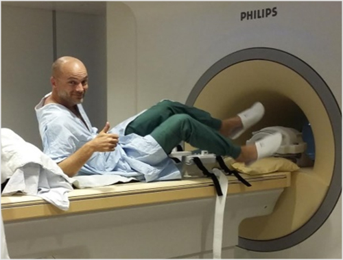
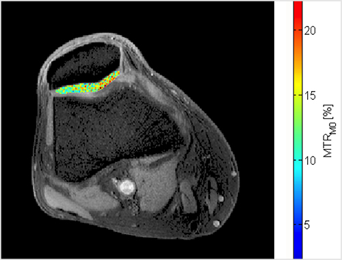
Structural imaging We use multiple cohort data sets with repeat magnetic resonance imaging and post processing of conventional radiographs to gain new knowledge of early stage osteoarthritis and its prediction.
Burden of disease Using population-based health care registries in Sweden covering in excess of 20 million person-years, we study the epidemiology and burden of musculoskeletal disease, including health economic aspects.
News
CHECK OUT THE ARTHRITIS PORTAL (SWE or ENG). Created by Lund University researchers, the arthritis portal is a platform where we share accurate and up-to-date information about osteoarthritis, including the latest development in research.
2026-02-16: We welcome new post doctoral student Isabel Mauricio Amado who will work with biomechanics and explant ex vivo studies.
2025-12-24: We at the Clinical epidemiology group wish you all a Merry Christmas and a Happy New Year! We look forward to 2026 and a new year of novel scientific discoveries.
2025-12-19: Just before Christmas orthopaedic surgeon Jamie Brown defended his PhD thesis "Acute knee injury with focus on ligaments other than the anterior cruciate", with Prof. Sebastian Kopf from the University Brandenburg an der Havel as thesis opponent. You can read more on his work here.
2025-09-12: A friday in September, orthopedist PhD student Anders Isacsson presented his thesis "On soft-tissue knee injures - epidemiology and outcome", with Prof. Jüri Kartus from the University of Gothenburg as thesis opponent. Congratulations to Anders for finally completing his thesis! You can read more on his work here.
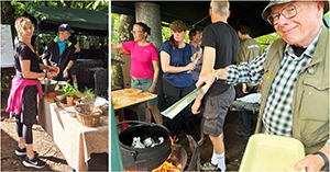
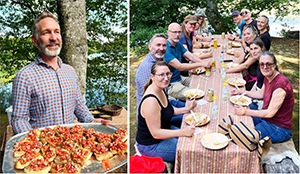
2025-09-04: This years annual kickoff took place at the wildlife camp in Häckeberga, where we enjoyed a great day outdoors, cooking our own lunch over fire and taking a lovely walk around the lake.
Starting the day with breakfast and coffee, and then the cooking begins!
Full activity, cooking all the food over fire!
Finally, we enjoyed all the wonderful food together.
2025-05-16: In May, clinical PhD student Fredrik Boric-Persson successfully defended his thesis: "Meniscus repair - Long-term gains with short-term challenges? We congratulate Fredrik and thank thesis opponent Professor Kristian Samuelsson from University of Gothenburg! You can read Fredrik's thesis here.
2025-04-27: This years OARSI congress took place in Incheon, South Korea, with Prof. Martin Englund as OARSI president. Here, PhD student Filippo Recenti got to present his research on the association between metabolic conditions and physical activity in osteoarthritis in an oral session (ABSTRACT), and Associate Prof. Andrea Dell'Isola presented his work on metabolic syndrome and osteoarthritis (abstract) and also got to present the "Epidemiology and Therapy" section in the session "Year in Review".
In addition, we presented several posters, with a selection listed here:
Amanda Sjögrenet al: Knee cartilage aging: histological analysis of donors without clinical knee disease aged 18 to 85 years (Abstract) Andrea Dell'Isolaet al: Does changing willingness for surgery translate to spared joint replacements? A 9-year longitudinal study on 55,000 individuals (Abstract) Karin Magnussonet al: Metformin and incident osteoarthritis: Causal inference from a co-twin control study (Abstract) Aleksandra Turkiewiczet al: The fallancy of responder analysis when studying treatments for osteoarthritis pain (Abstract)
Publications
Highlights
Association between ACL continuity on magnetic resonance imaging at 5 years after an acute ACL rupture and 11-Year outcomes: a secondary analysis from the KANON trial. Stephanie R Filbay, Frank Roemer, Ewa M Roos, Aleksandra Turkiewicz, Richard Frobell, L Stefan Lohmander, Martin Englund. The American Journal of Sports Medicine, 53(8):1893-1900, July 2025.
Background: Emerging evidence suggests that anterior cruciate ligament (ACL) ruptures can restore ACL fiber continuity. The relationship between ACL continuity on magnetic resonance imaging (MRI) (sign of ACL healing) and outcomes >5 years after an acute ACL rupture has not been investigated.
Purpose: This study aimed to (1) describe clinical outcomes and radiographic osteoarthritis (ROA) at 11 years based on ACL continuity status at 5 years and (2) investigate the relationship between 5-year ACL continuity status and 11-year Knee Injury and Osteoarthritis Outcome Score (KOOS4) scores.
Study design: Secondary analysis of KANON randomized controlled trial; Level of evidence, 3.
Methods: Overall, 105 of 121 (87%) active adults with acute ACL ruptures randomized to undergo initial exercise therapy and optional delayed ACL reconstruction (ACLR) or early ACLR and postoperative exercise therapy completed 11-year follow-up. MRI scans at 5 years were evaluated using the Anterior Cruciate Ligament OsteoArthritis Score (0-3), with grades 0 to 2 considered to represent "ACL continuity." Patient-reported outcomes (KOOS4, 36-Item Short Form Health Survey, Tegner Activity Scale, self-reported new knee injuries), knee laxity, and radiographic findings (tibiofemoral and/or patellofemoral ROA) were assessed at 11 years. The relationship between 5-year ACL continuity and 11-year KOOS4 scores (0-100) was examined using linear regression, adjusted for age, sex, smoking, and baseline KOOS4 scores.
Results: Of patients managed nonsurgically, 58% (n = 14) had ACL continuity and 42% (n = 10) had ACL discontinuity at 5 years. Analyses suggest that ACL continuity was associated with worse 11-year KOOS4 scores compared with delayed ACLR (adjusted mean difference, -20.2 [95% CI, -31.9 to -8.6]) and early ACLR (adjusted mean difference, -15.5 [95% CI, -26.4 to -4.7]) as well as similar or worse KOOS4 scores compared with ACL discontinuity (adjusted mean difference, -11.4 [95% CI, -26.5 to 3.6]). The proportion of patients with tibiofemoral ROA ranged from 14% (ACL continuity) to 23% (delayed ACLR), and the proportion of patients with patellofemoral ROA ranged from 11% (ACL discontinuity) to 41% (early ACLR).
Conclusion: ACL continuity on 5-year MRI may be associated with worse patient-reported outcomes at 11 years after an ACL injury compared with early or delayed ACLR.
Regression to the mean for physical function and quality of life in clinical trials for symptomatic knee osteoarthritis. Martin Englund, Aleksandra Turkiewicz. Osteoarthritis and Cartilage, 33(3):391-395, March 2025.
Objective: To estimate the size of regression to the mean (RTM) for common patient-reported outcomes in trials for knee osteoarthritis (OA).
Design: Longitudinal cohort study; we included participants of the Osteoarthritis Initiative who fulfilled typical inclusion criteria for enrolment in a trial. These included: age 40-79 years, symptomatic knee OA, Kellgren-Lawrence grade 2-3, use of pain medication more than half the days of a month in past 12 months, numerical rating scale pain of 4 to 9. We studied observed changes in WOMAC physical function and KOOS quality of life (QOL).
Results: We identified 547 subjects who fulfilled inclusion criteria on at least one annual follow-up between year 1 and year 8. The mean level of physical function and QOL at each follow-up time point was similar, about 18 and about 51, respectively. However, at the time of theoretical inclusion in a trial, the mean levels in the same subjects were 23 and 43, respectively (both worse scores). The mean improvement in physical function between inclusion and 1 and 2 years later, respectively, was 2.5 (95% confidence interval 1.7 to 3.2) and 3.1 (2.3 to 3.8). The corresponding improvement in QOL was 2.7 (1.7 to 3.7) and 4.2 (3.1 to 5.3).
Conclusion: RTM in trials for knee OA is likely to explain improvement in physical function and QOL, not only in knee pain. RTM often misleads investigators to overinterpret effectiveness as RTM neither represents improvement from the intervention nor placebo effect from the intervention and its context.
Altered co-expression patterns of synovial fluid proteins related to the immune system and extracellular matrix organization in late stage OA, compared to non-OA controls. Jenny Lönsjö, Martin Rydén, Aleksandra Turkiewicz, Velocity Hughes, Jon Tjörnstrand, Patrik Önnerfjord, Martin Englund, Neserin Ali. Frontiers in Immunology, 16:1523103, June 2025.
Objective: Synovial fluid contains proteins that may have been released from surrounding tissues, our aim was to gain new insights into the proteomic profiles of human synovial fluid in knees with and without osteoarthritis (OA).
Methods: We used synovial fluid from 11 patients with end-stage medial compartment knee OA, aspirated during total knee replacement, and from 13 deceased donors who had no prior history of knee OA (healthy controls). These samples were analyzed using high-multiplex immunoassays Olink®. The differential expression of proteins between the groups was analyzed using a linear mixed effects model. The linear associations between pairs of protein expressions were estimated with a linear regression model.
Results: We found that almost half of the detected proteins were differentially expressed between the OA and non-OA controls. The proteins that were most elevated in the OA group compared to controls were tartrate-resistant acid phosphatase type 5 (fold change 10.6, 95% CI [6.6-17.0]), plasminogen activator inhibitor 1 (5.0 [3.1, 8.0]), coagulation factor XI (4.3 [2.6-6.8]) and urokinase-type plasminogen activator (4.3 [2.3-6.8]). The proteins with lower levels in OA compared to controls were fatty acid-binding protein, adipocyte (0.03 [0.02-0.05]), myocilin (0.05 [0.03-0.08]) and carbonic anhydrase 3 (0.14 [0.09-0.23]). The protein-protein co-expression analysis suggests an overall lower number of protein pairs that show co-expression in OA.
Conclusion: There is a substantial change in protein abundance in synovial fluid in end-stage knee OA, suggesting that global joint homeostasis is severely deranged. Our findings suggest altered co-expression between the immune response and extracellular matrix organization in end-stage knee OA, in comparison to non-OA controls.
Prevalence and classification of meniscal calcifications in the human knee. Bijay Ratna Shakya, Ville-Pauli Karjalainen, Iida Hellberg, Mikko AJ Finnilä, Khaled Elkhouly, Amanda Sjögren, Aleksandra Turkiewicz, Patrik Önnerfjord, Velocity Hughes, Jon Tjörnstrand, Martin Englund, Simo Saarakkala. Osteoarthritis and Cartilage, 32(11):1441-1451, November 2024.
Objective: To investigate the occurrence of meniscal calcifications in individuals with and without knee osteoarthritis (OA). Additionally, we aim to identify the specific types of calcifications: basic calcium phosphate (BCP) and calcium pyrophosphate dihydrate (CPP).
Method: We analyzed 82 meniscal posterior horn samples (medial and lateral) collected from 41 human subjects. Among them, 20 individuals underwent total knee replacement due to medial compartment OA, while 21 deceased donors had no known knee OA. The assessment of meniscal calcifications and Pauli's histopathological scoring was conducted using histological sections. Furthermore, adjacent sections underwent measurement using Raman spectroscopy to characterize BCP and CPP calcifications based on their distinct spectral fingerprints.
Results: All OA individuals exhibited calcifications in at least one meniscus, compared to 9.5% (95%CI 1%, 30%) of donors. Among 35 OA menisci with calcifications, 28(80%) had BCP, 5(14%) had CPP and 2(6%) had both types. In 4 donor menisci, 3(75%) had CPP while 1(25%) had both types. We estimated the association between Pauli score and presence of BCP in OA individuals, yielding an odds ratio of 2.1 (95%CI 0.8, 5.3) per 1 Pauli score. The association between Pauli score and presence of CPP (in whole study sample) seemed weaker, with odds ratio of 1.3 (95%CI 1.1, 1.7).
Conclusion: The presence of BCP was predominant in menisci of OA individuals, whereas CPP exhibited similar prevalence in individuals with and without OA. The formation of BCP crystals in menisci may represent an important and specific characteristic of OA disease process that warrants further attention.
Twenty-year trajectories of morbidity in individuals with and without osteoarthritis. Andrea Dell'Isola, Filippo Recenti, Martin Englund, Ali Kiadaliri. RMD open, 10(2):e004164, July 2024.
Objectives: To identify multimorbidity trajectories over 20 years among incident osteoarthritis (OA) individuals and OA-free matched references.
Methods: Cohort study using prospectively collected healthcare data from the Skåne region, Sweden (~1.4 million residents). We extracted diagnoses for OA and 67 common chronic conditions. We included individuals aged 40+ years on 31 December 2007, with incident OA between 2008 and 2009. We selected references without OA, matched on birth year, sex, and year of death or moving outside the region. We employed group-based trajectory modelling to capture morbidity count trajectories from 1998 to 2019. Individuals without any comorbidity were included as a reference group but were not included in the model.
Results: We identified 9846 OA cases (mean age: 65.9 (SD 11.7), female: 58%) and 9846 matched references. Among both cases and references, 1296 individuals did not develop chronic conditions (no-chronic-condition class). We identified four classes. At the study outset, all classes exhibited a low average number of chronic conditions (≤1). Class 1 had the slowest progression towards multimorbidity, which increased progressively in each class. Class 1 had the lowest count of chronic conditions at the end of the follow-up (mean: 2.9 (SD 1.7)), while class 4 had the highest (9.6 (2.6)). The presence of OA was associated with a 1.29 (1.12, 1.48) adjusted relative risk of belonging to class 1 up to 2.45 (2.12, 2.83) for class 4.
Conclusions: Our findings suggest that individuals with OA face an almost threefold higher risk of developing severe multimorbidity.
Exploring the early molecular pathogenesis of osteoarthritis using differential network analysis of human synovial fluid. Martin Rydén, Amanda Sjögren, Patrik Önnerfjord, Aleksandra Turkiewicz, Jon Tjörnstrand, Martin Englund, Neserin Ali. Molecular & Cellular Proteomics, 23(6):100785, June 2024.
The molecular mechanisms that drive the onset and development of osteoarthritis (OA) remain largely unknown. In this exploratory study, we used a proteomic platform (SOMAscan assay) to measure the relative abundance of more than 6000 proteins in synovial fluid (SF) from knees of human donors with healthy or mildly degenerated tissues, and knees with late-stage OA from patients undergoing knee replacement surgery. Using a linear mixed effects model, we estimated the differential abundance of 6251 proteins between the three groups. We found 583 proteins upregulated in the late-stage OA, including MMP1, collagenase 3 and interleukin-6. Further, we selected 760 proteins (800 aptamers) based on absolute fold changes between the healthy and mild degeneration groups. To those, we applied Gaussian Graphical Models (GGMs) to analyze the conditional dependence of proteins and to identify key proteins and subnetworks involved in early OA pathogenesis. After regularization and stability selection, we identified 102 proteins involved in GGM networks. Notably, network complexity was lost in the protein graph for mild degeneration when compared to controls, suggesting a disruption in the regular protein interplay. Furthermore, among our main findings were several downregulated (in mild degeneration versus healthy) proteins with unique interactions in the healthy group, one of which, SLCO5A1, has not previously been associated with OA. Our results suggest that this protein is important for healthy joint function. Further, our data suggests that SF proteomics, combined with GGMs, can reveal novel insights into the molecular pathogenesis and identification of biomarker candidates for early-stage OA.
Genetic influence to osteoarthritis versus other rheumatic diseases. Karin Magnusson, Alexandra Turkiewicz, Martin Rydén, Martin Englund. Arthritis & Rheumatology, 76(2):206-215, February 2024.
Objective: To compare the genetic contribution to osteoarthritis (OA) vs other rheumatic/musculoskeletal diseases (RMDs) in the same population, and 2) to explore the role for any shared genetics between OA and other RMDs.
Methods: In 59 970 Swedish twins aged 35 years or older, we estimated the heritability (the variance explained by genetic factors) to OA in peripheral joints, back and neck pain, shoulder pain (adhesive capsulitis, impingement syndrome, etc), rheumatoid arthritis (RA), spondyloarthritis and psoriatic arthritis (SpA/PSA), myalgia and osteoporosis diagnosed in specialist and inpatient care. We also studied how much of covariance between OA and each of the RMDs could be explained by genetics, by studying phenotypic correlations in bivariate classical twin models.
Results: Any-site OA and hip OA (50% and 64%) were among the most heritable RMDs (as compared to 23% for fibromyalgia (lowest) and 63% for spondylarthritis (highest)). The highest phenotypic correlations were between OA (any joint site) and shoulder pain in the same individual (r=0.33, 95% CI=0.31-0.35), of which 70% (52-88) could be explained by shared genetics. The phenotypic correlation between OA and back/neck pain was r=0.25, with 25%-75% explained by genetics. Phenotypic correlations between OA and each of the other RMDs were lower (r~0.1 to r~0.2) with inconclusive sources of variation.
Conclusion: OA has relatively large heritability as compared to other RMDs. The co-existence of OA and shoulder pain, as well as back pain, was common and could often be explained by genetic factors. Findings imply similar etiologies of OA and several pain conditions.
Identification and quantification of degradome components in human synovial fluid reveals an increased proteolytic activity in knee osteoarthritis patients vs control. Martin Rydén, Alexandra Turkiewicz, Patrik Önnerfjord, Jon Tjörnstrand, Martin Englund, Neserin Ali. Proteomics, 23(15):e2300040, August 2023.
Synovial fluid (SF) may contain cleavage products of proteolytic activities. Our aim was to characterize the degradome through analysis of proteolytic activity and differential abundance of these components in a peptidomic analysis of SF in knee osteoarthritis (OA) patients versus controls (n = 23). SF samples from end-stage knee osteoarthritis patients undergoing total knee replacement surgery and controls, that is, deceased donors without known knee disease were previously run using liquid chromatography mass spectrometry (LC-MS). This data was used to perform new database searches generating results for non-tryptic and semi-tryptic peptides for studies of degradomics in OA. We used linear mixed models to estimate differences in peptide-level expression between the two groups. Known proteolytic events (from the MEROPS peptidase database) were mapped to the dataset, allowing the identification of potential proteases and which substrates they cleave. We also developed a peptide-centric R tool, proteasy, which facilitates analyses that involve retrieval and mapping of proteolytic events. We identified 429 differentially abundant peptides. We found that the increased abundance of cleaved APOA1 peptides is likely a consequence of enzymatic degradation by metalloproteinases and chymase. We identified metalloproteinase, chymase, and cathepsins as the main proteolytic actors. The analysis indicated increased activity of these proteases irrespective of their abundance.
Pain in clinical trials for knee osteoarthritis: estimation of regression to the mean. Martin Englund, Aleksandra Turkiewicz. The Lancet Rheumatology, 5(6):309-311, April 2023.
Evidence of ACL healing on MRI following ACL rupture treated with rehabilitation alone may be associated with better patient-reported outcomes: a secondary analysis from the KANON trial. Stephanie Filbay, Frank Roemer, Stefan Lohmander, Aleksandra Turkiewicz, Ewa Roos, Richard Frobell, Martin Englund. British Journal of Sports Medicine, 57(2):91-98, November 2022.
Objectives: Evaluate the natural course of anterior cruciate ligament (ACL) healing on MRI within 5 years of acute ACL rupture and compare 2-year and 5-year outcomes based on healing status and treatment group.
Methods: Secondary analysis of 120 Knee Anterior Cruciate Ligament Nonsurgical vs Surgical Treatment (KANON) trial participants randomised to rehabilitation and optional delayed ACL reconstruction (ACLR) or early ACLR and rehabilitation. ACL continuity on MRI (Anterior Cruciate Ligament OsteoArthritis Score 0-2) was considered evidence of ACL healing. Outcomes included Knee Injury and Osteoarthritis Outcome Score (KOOS), KOOS patient acceptable symptomatic state (PASS) and treatment failure criteria. Linear mixed models were used to estimate adjusted mean differences (95% CIs) in patient-reported sport and recreational function (KOOS-Sport/Rec) and quality of life (KOOS-QOL) at 2 and 5 years, between participants with MRI evidence of ACL healing and those who had (1) no evidence of ACL healing, (2) delayed ACLR or (3) early ACLR.
Results: MRI evidence of ACL healing at 2-year follow-up was observed in 16 of 54 (30%, 95% CI 19 to 43%) participants randomised to optional delayed ACLR. Excluding participants who had delayed ACLR, 16 of 30 (53%, 36-70%) participants managed with rehabilitation-alone displayed MRI evidence of ACL healing. Two-year outcomes were better in the healed ACL group (n=16) compared with the non-healed (n=14) (mean difference (95% CI) KOOS-Sport/Rec: 25.1 (8.6-41.5); KOOS-QOL: 27.5 (13.2-41.8)), delayed ACLR (n=24) (KOOS-Sport/Rec: 24.9 (10.2-39.6); KOOS-QOL: 18.1 (5.4-30.8)) and early ACLR (n=62) (KOOS-Sport/Rec: 17.4 (4.1-30.7); KOOS-QOL: 11.4 (0.0-22.9)) groups. Five-year KOOS-QOL was better in the healed versus non-healed group (25.3 (9.4-41.2)). Of participants with MRI evidence of ACL healing, 63-94% met the PASS criteria for each KOOS subscale, compared with 29-61% in the non-healed or reconstructed groups.
Conclusions: MRI appearance of ACL healing after ACL rupture occurred in one in three adults randomised to initial rehabilitation and one in two who did not cross-over to delayed ACLR and was associated with favourable outcomes. The potential for spontaneous healing of the ACL to facilitate better clinical outcomes may be greater than previously considered.